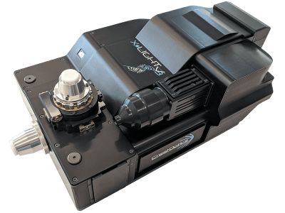
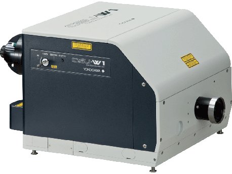
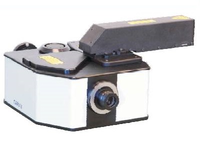
Crisel Electrooptical Systems & Technology logo

The CARV II™ Confocal Imager from Crisel Electrooptical Systems & Technology s.r.l.

The CARV II™ Confocal Imager is a fully automated, full-spectrum spinning disk confocal imager that attaches to most major models of inverted fluorescence microscope. It is ideal for live cell imaging in biological applications.

It offers the following:

  • Automated dichroic wheel and filter wheels
  • Camera placed in direct emission light path
  • FRAP iris
  • Manual control via keypad as well as automated software control
  • Easier alignment
  • Higher-throughput Semrock filters

Technical specifications are available.

CARV II System

CARV II Features and Benefits

Multipoint Confocal Scanning

The CARV II Confocal Imager module utilizes a Nipkow spinning disk which contains multiple sets of spirally arranged pinholes placed in the image plane of the objective lens. The column of excitation light is split into 1,000 beams to simultaneously scan the entire field once every millisecond, effectively creating a full image of the focal plane in real time. Emitted light is collected and imaged using a high‐resolution and high‐quantum efficiency CCD camera.

Below is a schematic representation of confocal imaging. Panel 1 is a simplified diagram of the emission path of a confocal imaging platform. Panel 2 shows the manner of application of the focused light to the specimen by either laser raster scanning (1) or through a Nipkow disk system such as the CARV II (2).


Panel 1 shows that a confocal microscope keeps out‐of‐focus light from reaching the detector.

Panel 2 illustrates the ability of the Nipkow disk to pass light during one of the scans available with each second. At a given moment, the sample is illuminated with 1,000 discrete points of light over a circular area 13 mm in diameter.

As the disk spins, the holes allow the passage of light across the entire field. The result is a full image of the focal plane. Because passage across the entire field occurs once every millisecond, creation of the full image takes place in real time.

The field of view available with cameras used with the CARV II is well suited to biological imaging. At 60× magnification, a CCD camera with a ½‐inch chip images an area 150 µm × 100 µm in size.

The spinning disk approach offers the ability to monitor rapidly occurring events within living cells without compromising resolution and it delivers substantial reduction of photobleaching and phototoxicity due to high‐frequency low‐intensity illumination.

Direct Viewing and Imaging of Confocal and Wide Field

The CARV II Confocal Imager permits direct viewing of confocal images through a binocular eyepiece, through the camera, or both. It is the only pinhole spinning disk fluorescence confocal system which allows the user to quickly switch from confocal to wide-field viewing or recording.

Sea Urchin Egg (Tubulin). Dr. George von Dassow, University of Washington, Friday Harbor, WA

Full Spectrum Confocal

The CARV II Confocal Imager uses a Mercury/Metal Halide Arc lamp as an illumination source. This allows full spectrum (360nm-700nm), real-time confocal imaging. In combination with the vast array of commercially available filter sets, any fluorescent marker can be confocally imaged at a fraction of the cost of laser‐based systems.

Automated filter selection

Automation of internal eight-position excitation, five-position dichroic and eight-position emission filter wheels allows fast multi-dimensional confocal imaging. It reduces the reliance on multi-band pass filter sets, allowing maximum light throughput and fast sequential imaging of multiple fluorescent probes in a given sample.

The automated wheels can handle as many as five fluorescent probes, and can handle still more with the use of suitable dichroic filters.

Fluorescence Recovery After Photobleaching (FRAP) capabilities

A FRAP iris at the same focal plane as the confocal disk creates an adjustable rectangular aperture on the image. Passage of high‐intensity Hg/metal halide light through this slit enables controlled photo-bleaching of part of the sample followed by fluorescence recovery recording.

Microscope Compatibility

The CARV II Confocal Imager unit can be configured for use with most inverted fluorescence microscopes.

Application-specific Cameras

A wide selection of high-end cooled and non-cooled CCD cameras with a combination of 12-16 bit information, fast readout, high quantum efficiencies and small pixel sizes produces images at a high resolution and high signal-to-noise ratio.

3‐D Software Option

A range of state of the art 3‐D software packages can be used for acquisition and analysis of confocal images.

CARV II Specifications

Confocal scanner Nipkow spinning disk (pinholes)
Disk scan rate 1,000 scans per second
Pinhole diameter 70 µm
Spectral transmission 360‒700 nm
Z-resolution 0.5 µm (PSF); 100× PlanApo 1.4NA
Illumination source 120 W Hg/metal halide (1200 hr)
Internal excitation changer Automated eight‐position wheel (25 mm)
Internal dichroic changer Automated five‐position wheel (25.7 × 36 mm)
Internal emission changer Automated eight‐position wheel (25 mm)
Filter sets provided DAPI, E-GFP and Texas Red from Semrock Inc. BrightLine™ series
Operation mode Automated confocal, wide field, bright field
Observation Direct confocal binocular viewing or camera port
Detector compatibility Suitable CCD cameras from major makers such as:
  Andor Corporation
  The Cooke Corporation
  Photometrics
  Hamamatsu Photonics
  QImaging Corporation
Microscope compatibility Most inverted fluorescence microscopes with 100% camera port
FRAP Aperture control by hand via touchpad, by computer via RS‐232 serial interface
System control By hand via touchpad, by computer via RS‐232 serial interface
Software drivers Image-Pro Plus®, IPLab™, iVision‐Mac™, MetaFluor®, MetaMorph®. RS‐232 serial command set available
Size 11(W) × 15.5(L) × 6(H) inches 27.94(W) × 39.37(L) × 15.24(H) cm
Weight 14.5 lbs / 6.6 kg
Power 100 – 124V AC /12V DC

Applications

3D Reconstruction

CARV II System

Maximum projection and orthogonal view created from confocal sections through fixed and stained sea urchin embryo. Tubulin (green) and Actin (red).

60x 1.4 NA Nikon with CoolSNAP HQ camera.

Dr. George von Dassow, University of Washington, Friday Harbor, WA.

Full-spectrum, High-resolution Imaging

Stack projection of HeLa cells with Qdot™ Conjugates.

Nuclei—Qdot® 655 Goat anti-Mouse IgG Conjugate.

Golgi—Qdot® 585 Goat anti-Rabbit IgG conjugate.

Microtubules—Qdot® 525 Steptavidin Conjugate.

Quantum Dots/Invitrogen Corporation, Carlsbad, CA

Stack projection of mouse intestine section labeled with Alexa Fluor® 350 WGA, Alexa Fluor® 568 phalloidin, and SYTOX® Green.

Molecular Probes/Invitrogen, Eugene, OR

Alexa Fluor® and SYTOX® are registered trademarks of Molecular Probes, Inc, Eugene, OR.

IPLab is a trademark of Becton, Dickinson and Company, Franklin Lakes, NJ.

iVision‐Mac is a trademark of BioVision Technologies, Inc, Exton, PA.

MetaFluor and MetaMorph are registered trademarks of Molecular Devices, Inc., Sunnyvale, CA.

Image-Pro Plus is a registered trademark of Media Cybernetics, Inc., Bethesda, MD.

Qdot® is a registered trademark of the Quantun Dot Corporation, Hayward, CA.

BrightLine is a registered trademark of Semrock, Inc, Rochester, NY.