3D Cell Explorer
Tomographic Microscope from Nanolive SA
A revolutionary Tomographic Microscope for instant looks inside living cells in 3D
The 3D Cell Explorer is a high‐speed, high‐resolution and non‐invasive microscope that can look deep inside biological systems. This allows you to record stunning three‐dimensional images of entire cells and tissue slices in just seconds and with a higher resolution than any conventional microscope on the market. It is the result of years of hard work, innovation and an obsession to detail. With the 3D Cell Explorer, researchers, students and medical doctors can directly experience what happens inside the living cell in real time!
After you place the cellular specimen on the instrument, the 3D Cell Explorer performs a three‐dimensional acquisition every three seconds. Without invasive labeling of the specimen, you can observe your live cells as long as you wish, in a quantitative manner, based on the cells’ own physical properties (refractive index and refractive index gradient).
A Self-Adjusting, Holographic & Tomographic Microscope
The 3D Cell Explorer is a non‐invasive microscope that can image cells and tissues in vitro instantly in three dimensions without any stain or label. Through a combination of holography and rotational scanning the system measures precisely the four‐dimensional distribution of the physical refractive index within the cell. It is the result of years of work and development.
The sample is positioned between a high‐numerical‐aperture air objective below the sample and a rotational illumination arm above. Green light (520 nm) from a laser diode is split into sample and reference beams. The sample is illuminated with a laser beam inclined at 45° which rotates around the sample 360°. A series of holograms is recorded on a digital camera by combining the beam that has passed through the sample with the reference beam.
| Core characteristics and features of the 3D Cell Explorer hardware | |
|---|---|
| Unibody Construction | Nanolive has reduced structural complexity to one unique basic component – a unibody industrially derived from a single piece of aluminum. Thanks to this design, the 3D Cell Explorer is thin, light, simple, and stable. |
| Self-Adjusting Operation | The optics of the 3D Cell Explorer microscope self-calibrate for varying sample conditions (e.g. media evaporation, changes in Refractive Index). This achieves optimal results for every measurement. |
| Low-Power Laser | The 3D Cell Explorer uses a low-power laser (Laser class 1: 0.2 mW/mm2. No goggles are required. |
| Fast Acquisition | A full three‐dimensional image of your cell is displayed on your monitor within three seconds. |
| Long-term Observation | The microscope stage is perfectly compatible with classical top stage incubators. You are able to observe your cells’ behavior in their natural environment (5% CO2 and 37°C). |
| Technical Specifications | |
|---|---|
| Microscope Objective | Air 60× magnification with low-power laser |
| λ=520 nm | |
| Sample exposure 0.2mW/mm2 | |
| Resolution | Δxy = 200nm |
| Δz = 400nm | |
| Field of View | ca. 90 µm |
| Depth of Field | ca. 30 µm |
| Tomography Field Rate | 0.5 frames per second |
| three‐dimensional images | |
| full self-adjustment | |
| Sample stage ease of access | 60 mm of free access to the sample stage for sample manipulation |
Software
An intuitive and comprehensive tool for exploration of your cell data
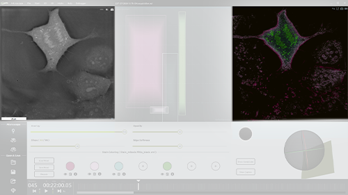
STEVE is the 3D Cell Explorer’s software counterpart. It processes the series of holograms captured by the hardware to create high-resolution images of each plane in the sample, employing a synthetic aperture and multiple-viewpoint-holographic methods to improve image resolution. STEVE’s intuitive interface provides control of the microscope, allows exploration of data using interactive digital staining and even performs quantitative analysis of measurements. STEVE runs smoothly, even during acquisition - by design.
Computational Imaging
The microscope registers one hundred holograms per rotation. Nanolive’s STEVE software processes the raw hologram data using complex deconvolution algorithms. STEVE also corrects for many imaging errors that would require extremely expensive optical components and ultra-precise alignment. The end result is a comprehensible 96-image Z-stack of the specimen displayed every three seconds.
- High-speed scanning: 20,000 points per second;
- Simple auto-calibration procedure;
- No optical switching;
- Diffraction-limited laser positioning;
- 3-D targeting;
- Streaming capabilities;
- Visible-light, UV, and IR light range;

Real‐Time
STEVE allows for the acquisition and processing of cell data in real time.
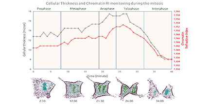
- One three‐dimensional image of the cell appears on the monitor every three seconds.
- One can follow the cell’s kinetics and cell’s dynamics live.
- Verification of microscope alignment is instananeous. Self‐calibration is performed when necessary.
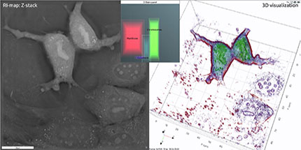
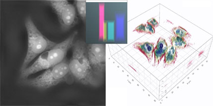
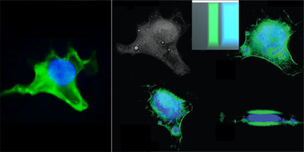
Digital Stain
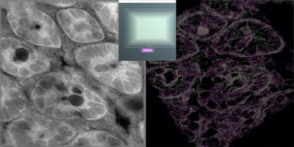
Explore your data in three‐dimensional view using our interactive digital stains and even perform quantitative analysis on your measurements.
- Quantitative staining based on physical markers (Refractive Index and Refractive Index Gradient)
- Manage your digital stains (create, edit, enable/disable, delete, save)
- Compare your results with other technologies, identify the exact Refractive Index range of your region of interest
- Export digital stains for analysis and comparison using other software

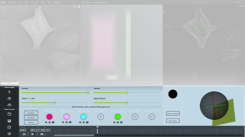
Interactive GUI
STEVE’s intuitive graphical user interface has been specifically conceived for ease of use and for fast learning. All options and commands necessary for acquiring and staining images are displayed on the main screen.
Simple and Advanced Microscope Control
- Single Shot acquisition
- Configurable time-lapse acquisition
- Object selection using auxiliary bright field mode
- Fully‐automated microscope calibration
- 3-D targeting
- Streaming capabilities
- Visible light, UV, and IR light range
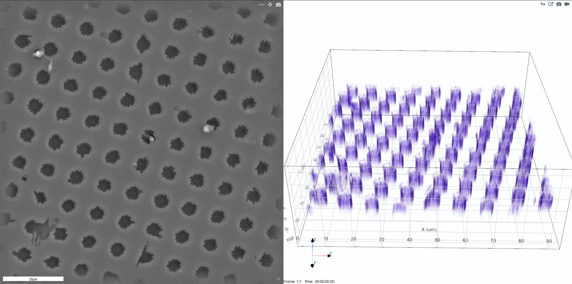
Comprehensive Visualization Options
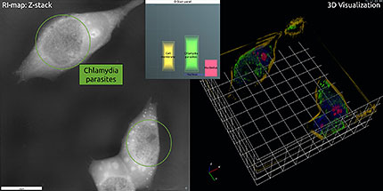
- Two visualization modes: Control mode and Viewer mode
- 2-D slice per slice viewer (refractive index and stained data combined)
- 3-D experience/scientific viewer
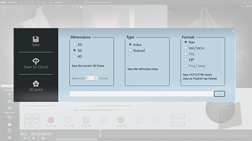
Multiple Data Export Options
- Classical Formats: Raw, TIFF, OBJ, PNG/JPEG
- Compressed proprietary formats: VOL/VOLX
- Screenshot capture for 2-D and 3-D viewers
- Video capture of the 3D viewer in AVI format
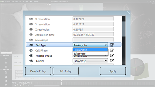
Data Annotation System
- Keep track of your data
- Classify your data based on different info tags
- Create customized info tags